
Introduction
The ocular drug development landscape is rapidly expanding, encompassing small molecules, biologics, gene and cell therapies, and novel drug delivery platforms. Increasingly, ocular delivery routes such as intravitreal, subconjunctival, and periocular administration are being used to target the posterior segment of the eye and support the development of injectable and implantable prolonged-action dosage forms.
Nonclinical development of ocular drugs involves pharmacology, pharmacokinetics, and toxicity studies conducted in accordance with the ICH M3 guideline and ICH S4 guidance. Despite this, there is currently no dedicated regulatory guideline specifically addressing safety evaluation through ocular delivery.
Ocular toxicity studies are unique because the test article is administered directly to the target organ, the eye. This direct exposure differentiates them from systemic toxicity studies and necessitates specialized study design, species selection, dosing considerations, and comprehensive ophthalmic evaluations.
Modes of Ocular Drug Delivery and Their Toxicological Relevance
Several routes are employed for ocular drug delivery depending on therapeutic intent:
- Topical administration (eye drops): Commonly used in toxicity studies targeting anterior segment diseases.
- Intravitreal (IVT) injection
- Subconjunctival injection
- Intracameral (IC) administration: Frequently used for antibiotics delivered into the anterior chamber.
- Subretinal administration: Used mainly in gene therapy to target retinal cells.
- Periocular administration: Takes advantage of scleral permeability and is useful for sustained-release drugs, corticosteroids, and anesthetics.
- Suprachoroidal (SC) delivery: A minimally invasive microneedle-based technique delivering drugs into the space between the sclera and choroid.
It is important to note that not all routes are feasible or appropriate in laboratory animal models, and route selection must be justified scientifically.
Animal Species Selection for Ocular Toxicity Studies
While the ICH M3 guideline recommends two species (one rodent and one non-rodent) for general toxicity studies, ocular toxicity testing follows a different paradigm:
- Rabbits are used in approximately 90% of ocular drug development programs as the first species.
- Cynomolgus monkeys are the next most frequently used species, followed by dogs.
- Species selection is performed case-by-case, with scientific justification.
Minipigs as Emerging Models
Minipigs offer several advantages:
- Inability to regenerate corneal endothelium, similar to humans
- Eyelid skin anatomy and physiology closely resemble humans
- Comparable cone-to-rod ratio and large vitreous volume
- Overall ocular anatomy and physiology closer to humans than rabbits
Regulatory Expectations Following Ocular Exposure
For first-in-human studies (FIHS):
- Two species (usually non-rodents) via the ocular route are generally required.
- A single species may suffice with strong scientific rationale or supporting nonclinical/clinical data.
Key Regulatory Considerations:
- Ocular pharmacokinetics to assess absorption and distribution in ocular compartments (typically one species).
- Ocular tolerability studies conducted before pivotal repeat-dose studies.
- Evaluation of physicochemical properties, structure-activity relationships, and in vitro irritancy.
- European authorities recommend single-dose rabbit tolerance studies with slit-lamp biomicroscopy.
Pivotal Ocular Toxicity Evaluations Include:
- Biomicroscopy (anterior segment)
- Indirect ophthalmoscopy (posterior segment)
- Tonometry (intraocular pressure)
- Electroretinography (ERG) when retinal exposure occurs
- Histopathology
Systemic toxicokinetics are usually conducted, whereas ocular toxicokinetics are rarely feasible due to tissue limitations. Systemic toxicity evaluation via an alternative route is typically expected prior to FIHS.
Design Considerations for Ocular Toxicity Studies
Study design depends on:
- Clinical route and indication
- Pharmacokinetics and release characteristics
- Treatment frequency and duration
- Patient population (age, sex)
- Whether the product is a new chemical entity (NCE) or reformulation
Standard in-life parameters assessed include:
- Clinical observations
- Body weight and food consumption
- Clinical pathology
- Plasma bioanalysis for systemic exposure
For gene therapy ocular products, biodistribution studies using qPCR are critical to assess vector spread in ocular and systemic tissues.
Specialized Parameters in Ocular Toxicity Evaluation
Ophthalmic Assessments
- Biomicroscopy: Detailed evaluation of anterior ocular structures
- Indirect ophthalmoscopy: Preferred method for posterior segment evaluation
- Intraocular pressure (IOP): Measured using tonometry
- Electroretinography (ERG): Detects functional retinal changes
- Fluorescein staining: Assesses corneal epithelial integrity
- Biodistribution studies: Across ocular tissues such as cornea, retina, vitreous, and optic nerve
Additional imaging tools may include OCT, fundus photography, and pachymetry, depending on study objectives.
Immunogenicity Assessment
For biologic ocular drugs:
- Antidrug antibodies (ADA) in serum are evaluated to assess immune response and potential correlations with ocular inflammation.
For gene therapy products:
- ADA against viral vectors
- Cell-mediated immunity assessments using PBMCs
Gross Pathology and Histopathology
Ocular toxicity studies require meticulous tissue collection:
- Both treated and untreated eyes
- Eyelids, lacrimal glands, Harderian glands (if present)
- Optic nerve and associated muscles
Each eye undergoes:
- At least five H&E sections
- Evaluation of all ocular compartments, including species-specific macular regions
Systemic organ evaluation is typically conducted in control and high-dose groups for pivotal studies.
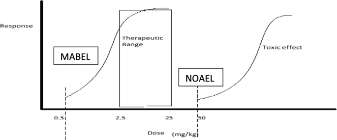
Data Interpretation and NOAEL Determination
A central objective of ocular toxicity studies is defining the No Observed Adverse Effect Level (NOAEL):
- NOAELs are determined using the complete dataset.
- Separate NOAELs are established for:
- Local ocular effects
- Systemic effects
Local and systemic toxicities may be independent, though systemic compromise can influence ophthalmic findings.
Conclusion
Translating animal ocular toxicity findings to humans requires careful consideration of:
- Species differences
- Safety margins
- Reversibility of effects
- Overall risk–benefit balance
The study director plays a crucial role in integrating findings related to the test article, vehicle, and procedure, and in coordinating interpretations among toxicologists, pathologists, and ophthalmologists to define adverse effects and establish scientifically sound NOAELs.